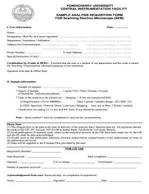
Scanning Electron Microscope SEM Pondicherry University Form


What is the Scanning Electron Microscope SEM Pondicherry University
The Scanning Electron Microscope (SEM) at Pondicherry University is a sophisticated instrument used for imaging and analyzing the surface structure of materials at a microscopic level. This technology allows researchers to obtain high-resolution images by scanning a focused beam of electrons across the sample surface. The SEM is particularly valuable in fields such as materials science, biology, and nanotechnology, providing insights into the morphology and composition of various materials.
How to use the Scanning Electron Microscope SEM Pondicherry University
Using the Scanning Electron Microscope involves several steps to ensure accurate results. First, samples must be prepared properly, which may include coating non-conductive materials with a thin layer of conductive material. Once prepared, users can load the samples into the SEM chamber. The microscope operates by adjusting the electron beam and detectors to capture detailed images. Proper training and adherence to safety protocols are essential for effective operation.
Steps to complete the Scanning Electron Microscope SEM Pondicherry University
Completing the process with the SEM includes the following steps:
- Prepare the sample by cleaning and, if necessary, coating it with a conductive layer.
- Load the sample into the SEM chamber, ensuring it is securely placed.
- Set the desired operating conditions, including voltage and beam current.
- Focus the electron beam on the sample and adjust the imaging parameters.
- Capture images and analyze the data as needed.
Legal use of the Scanning Electron Microscope SEM Pondicherry University
The legal use of the Scanning Electron Microscope at Pondicherry University is governed by institutional policies and relevant regulations regarding research and data handling. Users must ensure compliance with ethical standards, particularly when working with biological samples or sensitive materials. Additionally, proper documentation of research findings is essential for maintaining integrity and accountability in scientific research.
Key elements of the Scanning Electron Microscope SEM Pondicherry University
Key elements of the SEM include:
- Electron Source: The component that generates the electron beam.
- Sample Chamber: Where the sample is placed for imaging.
- Detectors: Devices that capture emitted signals from the sample.
- Control System: Software and hardware that manage the microscope's settings and operations.
- Vacuum System: Maintains a vacuum environment to prevent electron scattering.
Examples of using the Scanning Electron Microscope SEM Pondicherry University
Examples of applications for the Scanning Electron Microscope at Pondicherry University include:
- Analyzing the surface morphology of nanomaterials.
- Examining biological specimens, such as cells and tissues, for structural studies.
- Investigating the properties of metals and alloys for engineering applications.
- Studying the effects of corrosion on various materials.
Quick guide on how to complete scanning electron microscope sem pondicherry university
Manage Scanning Electron Microscope SEM Pondicherry University with ease on any device
Digital document handling has become increasingly favored by companies and individuals alike. It offers an ideal environmentally friendly alternative to conventional printed and signed documents, allowing you to access the correct form and securely save it online. airSlate SignNow equips you with all the necessary tools to create, modify, and electronically sign your files swiftly and without delays. Manage Scanning Electron Microscope SEM Pondicherry University on any device using the airSlate SignNow apps for Android or iOS and enhance your document-centric workflows today.
How to modify and electronically sign Scanning Electron Microscope SEM Pondicherry University effortlessly
- Obtain Scanning Electron Microscope SEM Pondicherry University and then click Get Form to begin.
- Leverage the tools available to complete your form.
- Emphasize important sections of the documents or redact confidential information using the tools specifically provided by airSlate SignNow for that purpose.
- Create your electronic signature with the Sign feature, which takes just seconds and carries the same legal validity as a conventional handwritten signature.
- Review the details and then click the Done button to save your changes.
- Choose your preferred method for submitting your form—via email, text message (SMS), invitation link, or download it directly to your computer.
Eliminate the worry of lost or misplaced documents, tedious form searching, or mistakes that necessitate printing new copies. airSlate SignNow addresses all your document management needs with just a few clicks from any device of your choice. Edit and electronically sign Scanning Electron Microscope SEM Pondicherry University to maintain effective communication at every step of the document preparation journey with airSlate SignNow.
Create this form in 5 minutes or less
Create this form in 5 minutes!
How to create an eSignature for the scanning electron microscope sem pondicherry university
How to create an electronic signature for a PDF online
How to create an electronic signature for a PDF in Google Chrome
How to create an e-signature for signing PDFs in Gmail
How to create an e-signature right from your smartphone
How to create an e-signature for a PDF on iOS
How to create an e-signature for a PDF on Android
People also ask
-
What is a Scanning Electron Microscope SEM at Pondicherry University?
The Scanning Electron Microscope SEM at Pondicherry University is a state-of-the-art imaging tool that provides high-resolution images at the nanoscale level. It is widely used in various research fields, including materials science, biology, and nano-technology. This advanced equipment allows researchers to explore the microstructure of samples with exceptional detail.
-
How much does it cost to use the Scanning Electron Microscope SEM at Pondicherry University?
Pricing for using the Scanning Electron Microscope SEM at Pondicherry University varies based on the type of research and the time required for usage. Prospective customers are encouraged to contact the university's laboratory services for tailored pricing information. Generally, the SEM services are cost-effective and aimed at supporting academic and research endeavors.
-
What features does the Scanning Electron Microscope SEM at Pondicherry University offer?
The Scanning Electron Microscope SEM at Pondicherry University boasts features like high magnification, varying resolutions, and a range of detectors for material characterization. It also includes capabilities for elemental analysis using Energy Dispersive X-ray Spectroscopy (EDS). These features make it versatile for numerous applications in scientific research.
-
What are the benefits of using the Scanning Electron Microscope SEM at Pondicherry University?
Using the Scanning Electron Microscope SEM at Pondicherry University offers numerous benefits, including access to advanced imaging technologies that can enhance research outcomes. The ability to visualize samples at different magnifications aids in comprehensive analysis. Furthermore, the support from skilled technicians ensures effective use of the equipment for optimal results.
-
Is training available for using the Scanning Electron Microscope SEM at Pondicherry University?
Yes, Pondicherry University provides training sessions for users of the Scanning Electron Microscope SEM. The training covers operational protocols, safety measures, and data analysis techniques. These sessions are designed to empower users, ensuring they can utilize the SEM effectively for their research.
-
Can the Scanning Electron Microscope SEM at Pondicherry University be integrated with other laboratory instruments?
The Scanning Electron Microscope SEM at Pondicherry University can often be integrated with other analytical tools, such as X-Ray Diffraction (XRD) and Atomic Force Microscopy (AFM). This integration allows for a more comprehensive analysis of materials, enhancing research capabilities. Collaboration between different instruments can lead to richer data and improved findings.
-
What types of samples can be analyzed using the Scanning Electron Microscope SEM at Pondicherry University?
The Scanning Electron Microscope SEM at Pondicherry University can analyze a diverse range of samples, including metals, polymers, biological specimens, and nanomaterials. Its versatility makes it suitable for various applications across multiple scientific disciplines. However, users are advised to check sample preparation requirements specific to SEM analysis.
Get more for Scanning Electron Microscope SEM Pondicherry University
- Invitation to appear as a guest speaker event coordinator to guest form
- Form rev 811 form 35e price escalation addendum
- In a powerball lottery pool use our lottery pool agreement form
- Titles rules and regulations mississippi department of form
- Notice to lessorlessees intention not to renew or extend lease agreement form
- Demandfor address of party under statute or rule form
- Medical consent letter with enclosed form
- Request for address assignmentverification the city of san form
Find out other Scanning Electron Microscope SEM Pondicherry University
- Can I Electronic signature Hawaii Insurance PDF
- Help Me With Electronic signature Colorado High Tech Form
- How To Electronic signature Indiana Insurance Document
- Can I Electronic signature Virginia Education Word
- How To Electronic signature Louisiana Insurance Document
- Can I Electronic signature Florida High Tech Document
- Can I Electronic signature Minnesota Insurance PDF
- How Do I Electronic signature Minnesota Insurance Document
- How To Electronic signature Missouri Insurance Form
- How Can I Electronic signature New Jersey Insurance Document
- How To Electronic signature Indiana High Tech Document
- How Do I Electronic signature Indiana High Tech Document
- How Can I Electronic signature Ohio Insurance Document
- Can I Electronic signature South Dakota Insurance PPT
- How Can I Electronic signature Maine Lawers PPT
- How To Electronic signature Maine Lawers PPT
- Help Me With Electronic signature Minnesota Lawers PDF
- How To Electronic signature Ohio High Tech Presentation
- How Can I Electronic signature Alabama Legal PDF
- How To Electronic signature Alaska Legal Document