The Diagram Shows Some of the Cell Structures Involved in the Secretion of an Extracellular Enzyme Form


What is the diagram shows some of the cell structures involved in the secretion of an extracellular enzyme?
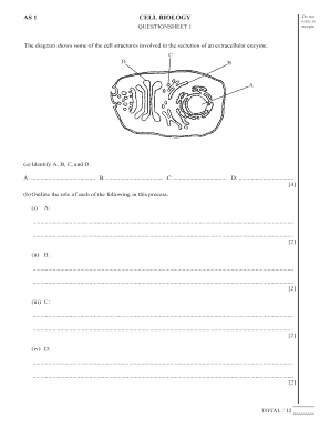
The diagram shows some of the cell structures involved in the secretion of an extracellular enzyme, illustrating the complex biological processes that occur within cells. This diagram typically highlights key components such as the endoplasmic reticulum, Golgi apparatus, and vesicles. Each structure plays a vital role in synthesizing, processing, and transporting enzymes outside the cell. Understanding these components can provide insight into how cells communicate and interact with their environment, as well as the mechanisms of enzyme secretion.
How to use the diagram shows some of the cell structures involved in the secretion of an extracellular enzyme
To effectively use the diagram shows some of the cell structures involved in the secretion of an extracellular enzyme, start by familiarizing yourself with each component depicted. Identify the endoplasmic reticulum, which is responsible for synthesizing proteins, and the Golgi apparatus, which modifies and packages these proteins for secretion. Pay attention to how vesicles transport enzymes to the cell membrane. This understanding can enhance your comprehension of cellular functions and the importance of enzyme secretion in various biological processes.
Steps to complete the diagram shows some of the cell structures involved in the secretion of an extracellular enzyme
Completing the diagram shows some of the cell structures involved in the secretion of an extracellular enzyme involves several steps. First, gather all necessary materials, including a clear reference image of the diagram. Next, label each structure accurately, ensuring that terms like 'endoplasmic reticulum' and 'Golgi apparatus' are clearly indicated. After labeling, review the connections between these structures, emphasizing their roles in enzyme secretion. Finally, ensure that the diagram is neat and legible, making it easier for others to understand the cellular processes involved.
Key elements of the diagram shows some of the cell structures involved in the secretion of an extracellular enzyme
Key elements of the diagram shows some of the cell structures involved in the secretion of an extracellular enzyme include the endoplasmic reticulum, Golgi apparatus, and vesicles. The endoplasmic reticulum is crucial for protein synthesis, while the Golgi apparatus is essential for processing and packaging these proteins. Vesicles transport the finished enzymes to the cell membrane for secretion. Understanding these elements helps clarify the pathway of enzyme secretion and the intricate workings of cellular machinery.
Legal use of the diagram shows some of the cell structures involved in the secretion of an extracellular enzyme
The legal use of the diagram shows some of the cell structures involved in the secretion of an extracellular enzyme pertains to its application in educational and research contexts. When utilizing this diagram, it is important to ensure that proper citations are provided if the image is sourced from published materials. Additionally, any modifications made to the diagram should be clearly indicated to maintain transparency in academic and professional settings.
Examples of using the diagram shows some of the cell structures involved in the secretion of an extracellular enzyme
Examples of using the diagram shows some of the cell structures involved in the secretion of an extracellular enzyme can be found in various educational settings. In biology classes, instructors may use the diagram to teach students about cellular functions and the significance of enzyme secretion. Researchers may reference the diagram in publications to illustrate findings related to enzyme activity or cellular processes. Additionally, the diagram can serve as a visual aid in presentations, enhancing audience understanding of complex biological mechanisms.
Quick guide on how to complete the diagram shows some of the cell structures involved in the secretion of an extracellular enzyme
Manage The Diagram Shows Some Of The Cell Structures Involved In The Secretion Of An Extracellular Enzyme seamlessly on any device
Digital document management has gained signNow traction among businesses and individuals. It offers an ideal eco-friendly substitute to conventional printed and signed documents, allowing you to locate the appropriate form and securely store it online. airSlate SignNow equips you with all the necessary tools to create, edit, and electronically sign your documents swiftly without interruptions. Handle The Diagram Shows Some Of The Cell Structures Involved In The Secretion Of An Extracellular Enzyme on any device using airSlate SignNow's Android or iOS applications and simplify any document-related task today.
How to edit and electronically sign The Diagram Shows Some Of The Cell Structures Involved In The Secretion Of An Extracellular Enzyme effortlessly
- Obtain The Diagram Shows Some Of The Cell Structures Involved In The Secretion Of An Extracellular Enzyme and click Get Form to begin.
- Make use of the tools we provide to complete your form.
- Highlight important sections of your documents or redact sensitive information with tools specifically offered by airSlate SignNow for that purpose.
- Generate your electronic signature with the Sign tool, which takes mere seconds and holds the same legal validity as a traditional ink signature.
- Review the details and click on the Done button to save your modifications.
- Select how you wish to share your form, via email, text message (SMS), or invitation link, or download it to your computer.
Eliminate concerns about lost or misplaced documents, exhausting form searches, or mistakes that necessitate printing new document copies. airSlate SignNow addresses all your document management requirements in just a few clicks from any device you prefer. Revise and electronically sign The Diagram Shows Some Of The Cell Structures Involved In The Secretion Of An Extracellular Enzyme to ensure excellent communication at any stage of the form preparation process with airSlate SignNow.
Create this form in 5 minutes or less
Create this form in 5 minutes!
How to create an eSignature for the the diagram shows some of the cell structures involved in the secretion of an extracellular enzyme
How to create an electronic signature for a PDF online
How to create an electronic signature for a PDF in Google Chrome
How to create an e-signature for signing PDFs in Gmail
How to create an e-signature right from your smartphone
How to create an e-signature for a PDF on iOS
How to create an e-signature for a PDF on Android
People also ask
-
What is airSlate SignNow and how does it relate to cell structures?
airSlate SignNow is a powerful eSignature and document management platform that enables businesses to send and eSign documents efficiently. While the platform itself does not directly relate to biological concepts, understanding how the diagram shows some of the cell structures involved in the secretion of an extracellular enzyme can help inform users about processes involved in efficiency and effectiveness in various fields.
-
How does the pricing for airSlate SignNow compare to other eSignature solutions?
airSlate SignNow offers competitive pricing plans that cater to businesses of all sizes. By providing a cost-effective solution for eSigning documents, it enhances workflow efficiency—similar to how the diagram shows some of the cell structures involved in the secretion of an extracellular enzyme, facilitating essential processes.
-
What features does airSlate SignNow offer?
airSlate SignNow includes features like bulk sending, customizable templates, and advanced document analytics. These capabilities ensure that users can efficiently manage their document workflows, paralleling how the diagram shows some of the cell structures involved in the secretion of an extracellular enzyme, emphasizing the importance of streamlined processes.
-
Can airSlate SignNow integrate with other business software?
Yes, airSlate SignNow seamlessly integrates with various business applications such as CRM systems and cloud storage solutions. This integration enhances productivity and workflow, akin to how the diagram shows some of the cell structures involved in the secretion of an extracellular enzyme, allowing for interconnected processes.
-
What are the benefits of using airSlate SignNow for my business?
Using airSlate SignNow streamlines document management, reduces turnaround times, and improves overall operational efficiency. This is similar to how the diagram shows some of the cell structures involved in the secretion of an extracellular enzyme, emphasizing the significance of optimized processes in achieving successful outcomes.
-
Is there a free trial available for airSlate SignNow?
Yes, airSlate SignNow offers a free trial that allows businesses to experience the platform's capabilities firsthand. During this trial, users will see how document workflows can be simplified, much like how the diagram shows some of the cell structures involved in the secretion of an extracellular enzyme, promoting efficiency in operations.
-
How secure is airSlate SignNow for signing confidential documents?
airSlate SignNow employs top-notch security measures, including encryption and compliance with global standards, to protect sensitive information. This level of security ensures that your documents remain confidential, similar to how the diagram shows some of the cell structures involved in the secretion of an extracellular enzyme, safeguarding critical processes.
Get more for The Diagram Shows Some Of The Cell Structures Involved In The Secretion Of An Extracellular Enzyme
- Information about the petitioner check if in military
- Application for nz citizenship adult form
- Page 1 of 7 wb 5 form
- Plaintiffsnameampaddresstel form
- Oklahoma statutes title 43 marriage and family form
- Motion to vacate mediated stipulated judgment form
- Department of the air forcecfetp 2a3x3b form
- Elder restraining order 569542565 form
Find out other The Diagram Shows Some Of The Cell Structures Involved In The Secretion Of An Extracellular Enzyme
- How To eSignature Colorado Sponsorship Proposal Template
- eSignature Alabama Distributor Agreement Template Secure
- eSignature California Distributor Agreement Template Later
- eSignature Vermont General Power of Attorney Template Easy
- eSignature Michigan Startup Cost Estimate Simple
- eSignature New Hampshire Invoice for Services (Standard Format) Computer
- eSignature Arkansas Non-Compete Agreement Later
- Can I eSignature Arizona Non-Compete Agreement
- How Do I eSignature New Jersey Non-Compete Agreement
- eSignature Tennessee Non-Compete Agreement Myself
- How To eSignature Colorado LLC Operating Agreement
- Help Me With eSignature North Carolina LLC Operating Agreement
- eSignature Oregon LLC Operating Agreement Online
- eSignature Wyoming LLC Operating Agreement Online
- eSignature Wyoming LLC Operating Agreement Computer
- eSignature Wyoming LLC Operating Agreement Later
- eSignature Wyoming LLC Operating Agreement Free
- How To eSignature Wyoming LLC Operating Agreement
- eSignature California Commercial Lease Agreement Template Myself
- eSignature California Commercial Lease Agreement Template Easy