The EMBO Journal vol.15 no.24 pp.7156-7167, 1996
The catalytic subunit of protein phosphatase 2A
associates with the translation termination factor
eRF1
I
I
I
1
Natasa Andjelkovic,
Stanislaw Zolnierowicz1,
Christine Van Hoof 2, Jozef Goris2 and
Brian A.Hemmings3
Friedrich Miescher-Institut, PO Box 2543, CH-4002 Basel,
Switzerland and 2Afdeling Biochemie, Faculteit der Geneeskunde,
Katholieke Universiteit te Leuven, Herestraat 49, 3000 Leuven,
Belgium
'Present address: Department of Biochemistry, Faculty of
Biotechnology, Medical University of Gdansk, Debinki 1,
80-211 Gdansk, Poland
3Corresponding author
By a number of criteria, we have demonstrated that
the translation termination factor eRFi (eukaryotic
release factor 1) associates with protein phosphatase
2A (PP2A). Trimeric PP2A1 was purified from rabbit
skeletal muscle using an affinity purification step. In
addition to the 36 kDa catalytic subunit (PP2Ac) and
established regulatory subunits of 65 kDa (PR65) and
55 kDa (PR55), purified preparations contained two
proteins with apparent Mrs of 54 and 55 kDa. Protein
microsequencing revealed that the 55 kDa component
is a novel protein, whereas the 54 kDa protein was
identified as eRF1, a protein that functions in translational termination as a polypeptide chain release factor.
Using the yeast two-hybrid system, human eRF1 was
shown to interact specifically with PP2Ac, but not with
the PR65 or PR55 subunits. By deletion analysis, the
binding domains were found to be located within the
50 N-terminal amino acids of PP2Ac, and between
amino acid residues 338 and 381 in the C-terminal
part of human eRF1. This association also occurs
in vivo, since PP2A can be co-immunoprecipitated with
eRF1 from mammalian cells. We observed a significant
increase in the amount of PP2A associated with the
polysomes when eRF1 was transiently expressed in
COS1 cells, and eRF1 immunoprecipitated from those
fractions contained associated PP2A. Since we did
not observe any dramatic effects of PP2A on the
polypeptide chain release activity of eRF1 (or vice
versa), we postulate that eRF1 also functions to recruit
PP2A into polysomes, thus bringing the phosphatase
into contact with putative targets among the components of the translational apparatus.
Keywords: eRFI/protein phosphatase 2A/signal
transduction/translational termination
Introduction
Protein phosphatase 2A (PP2A) is implicated in the
regulation of many cellular processes including metabolism, signal transduction, growth, development, cell cycle
progression and transformation (reviewed in Mumby and
Walter, 1993; Mayer-Jaekel and Hemmings, 1994). PP2A
encompasses a family of trimeric holoenzymes which
consist of a 36 kDa catalytic subunit (PP2Ac) bound to
the constant regulatory subunit of 65 kDa (PR65/A) which
then associate further with the third, variable regulatory
subunit. Several trimeric PP2A holoenzymes have been
purified which contain different variable subunits of either
54, 55, 72 or 74 kDa (reviewed in Kamibayashi and
Mumby, 1995; Wera and Hemmings, 1995).
As documented by in vitro reconstitution assays and
by analyzing yeast and Drosophila mutants deficient in
regulatory proteins, both the constant and variable subunits
are important for controlling PP2A activity and substrate
specificity (reviewed in Mayer-Jaekel and Hemmings,
1994; Wera and Hemmings, 1995). For instance, PP2A
activity from brain extracts of Drosophila aarl mutants,
in which the gene encoding PR55 is disrupted by Pelement insertion, is several fold lower towards histone
HI and caldesmon phosphorylated by p34cdc2 as compared
with wild-type flies (Mayer-Jaekel et al., 1994). In contrast,
phosphorylase phosphatase activity of PP2A is similar in
aarl and control flies. The variable regulatory subunits
also represent targets for potential second messengers and
viral proteins. Dobrowsky et al. (1993) demonstrated that
ceramide activates only trimeric PP2A containing the
PR55 subunit whereas the PP2Ac-PR65 dimer is unaffected. Recent data, however, show that neither the constant
nor variable regulatory subunits are required for ceramide
stimulation of PP2A activity, since both PP2Ac and
PP2Ac-PR65 dimer can be stimulated by ceramide in a
manner similar to that of the trimeric holoenzyme, suggesting that PP2Ac itself is a target of ceramide action
(Law and Rossie, 1995). Furthermore, PP2A associates
with transforming antigens of certain DNA tumor viruses,
such as polyomavirus small t and middle T, and SV40
small t (Pallas et al., 1990). It is believed that these
oncoproteins act to alter PP2A activity by displacing the
normal cellular variable regulatory subunits from the
trimeric holoenzyme. Some viral proteins interact only
with specific forms of PP2A holoenzymes, e.g. SV40
small t antigen is able to replace only the B subunit
(PR55), but not the B' subunit (PR61) from trimeric PP2A
(Sontag et al., 1994). It was also shown that adenovirus
E4orf4 binds to the trimeric PP2A holoenzyme that
contains PR55 (Kleinberger and Shenk, 1993). Taken
together, these examples illustrate that the activity of
PP2Ac is tightly controlled in vivo by regulatory proteins.
We developed a strategy for simultaneous purification
of different PP2A holoenzymes from rabbit skeletal muscle
in order to analyze their subunit structure further. This
approach resulted in the purification of two heterotrimeric
forms of PP2Ao containing different isoforms of a novel
type of variable regulatory subunit (termed PR61) that
7
6 Oxford University Press
7156
i.
I
�PP2A associates with eRFl
Homogenate
Acidification to pH 5.3
DE52 batch elution with 0.3M NaCI
30-50% (NHd SO4 precipitation
DEAE Sepharose
(0.05-0.6 M NaCI)
pool I
pool 3
pool 2
~~~~~~~~~~~~~I,
X
minohexyl
Sepharose
I
I
Poly-L-lysine Agarose
|
mnohexyl
|Spharose
Thiophosphorylase-a Sepharose
MonoQ FPLC
PP2A0
SDS-PAGE analysis of pool 2 (fractions eluted from
DEAE-Sepharose between 0.34 and 0.38 M NaCl) after the
thiophosphorylase a-Sepharose purification step revealed
that this preparation contained the 36 kDa catalytic and
65 kDa regulatory subunits and several proteins in the
range of 54-55 kDa (Figure 2B). Further fractionation by
MonoQ FPLC resulted in the separation of PP2A, (trimer
containing the 55 kDa regulatory subunit, PR55) and
PP2A2 (the dimeric form of the enzyme) from two proteins
of 54 and 55 kDa (Figure 2A and C). These two proteins
appear to be unrelated to PR55 since they did not crossreact with anti-PR55 antibodies (data not shown). In
addition, we identified free PR55cx in this preparation by
SDS-PAGE and immunoblot analysis (fractions 37 and
38). This suggests that the PP2A2 identified probably
results from the dissociation of PR55, and possibly the
54 and 55 kDa proteins, from the complex (see below).
PP2A1+
associated
proteins
1
PP2A2
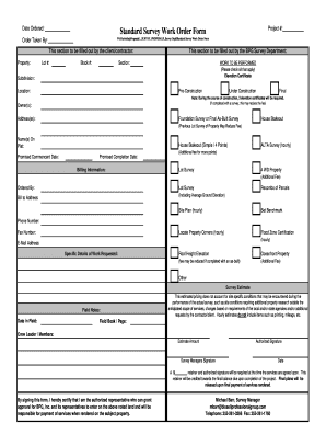
Fig. 1. Schematic outline of the purification protocol used to isolate
different PP2A holoenzymes from rabbit skeletal muscle.
probably function to target PP2A to nuclear substrates
(Tehrani et al., 1996; Zolnierowicz et al., 1996). We also
found two novel proteins of 54 and 55 kDa that apparently
co-purify with the trimeric PP2A1 holoenzyme following
affinity purification. Here we report the identification of
the 54 kDa protein as a member of the eRFI family of
proteins involved in termination of protein synthesis as
well as further examine the functional consequences of
its interaction with constituent components of the PP2A
holoenzyme.
Results
Co-purification of PP2A1 holoenzyme with two
proteins of 54 and 55 kDa
A modified protocol for PP2A purification from rabbit
skeletal muscle (see Figure 1 and Materials and methods)
was used to identify novel regulatory and/or associated
proteins. The partially purified material obtained from
DEAE-Sepharose pools 1, 2 and 3 was analyzed using
several antisera developed against the constituent subunits
of PP2A reported in earlier publications (Hendrix et al.,
1993a,b; Turowski et al., 1995). MonoQ FPLC of fractions
corresponding to pool 1 (eluted from DEAE-Sepharose
between 0.27 and 0.32 M NaCl) revealed the existence of
a trimeric PP2A holoenzyme containing 36 kDa catalytic
(PP2Ac) and 65 kDa regulatory (PR65) subunits and a
novel type of variable regulatory subunits with apparent
Mrs ranging from 56 to 61 kDa. This trimeric holoenzyme
apparently corresponds to PP2Ao (Zolnierowicz et al.,
1996) according to the classification established previously
by Tung et al. (1985).
54 kDa protein that co-purifies with PP2A1 is a
member of the eRF1 family of proteins with
polypeptide chain release factor activity
The 54 and 55 kDa proteins purified by MonoQ FPLC
chromatography were used to obtain protein sequence
data as described in Materials and methods. Sequences of
three tryptic peptides comprising 30 amino acids derived
from the 54 kDa protein were obtained: peptide 3, YFDEISQDTGK, peptide 9/10, ILYLTPEQEK and peptide 25/
26, S/GFGGIGGIL. Comparison of these sequences using
the FASTA program (Pearson and Lipman, 1988) revealed
76% homology to the predicted protein sequence encoded
by the Saccharomyces cerevisiae SUP45 gene (Breining
and Piepersberg, 1986). Human cDNAs corresponding to
this protein were isolated using a reverse transcriptionPCR approach (see Materials and methods). Among
several clones isolated from human fetal brain library and
analyzed by sequencing, only one, termed BBZ.eRFI-4b,
contained a full-length open reading frame (1311 bp) as
well as 231 bp of the 5'-non-coding region and -2.22 kb
of the 3'-non-coding region. This cDNA is identical to
the TB3-1 cDNA, originally identified by Grenett et al.
(1992) and resequenced by Frolova et al. (1994). cDNAs
encoding homologous proteins have been identified
recently from Arabidopsis thaliana (Quigley et al., 1994)
and Xenopus laevis (Tassan et al., 1993). The report by
Frolova et al. (1994) demonstrated that human and Xenopus proteins possess polypeptide chain release factor
activity and termed this factor eRFI. Sequence comparison
revealed that eRFI protein is highly conserved between
species, with yeast and human protein being 67.5%
identical (Figure 3).
Partial amino acid sequence analysis of seven peptides
comprising 71 amino acids derived from the 55 kDa
protein did not display any homologies to the sequences
present in currently available databases (GenBankTM,
release number 95). Currently we are attempting to isolate
cDNAs corresponding to this protein to establish its
relationship to PP2A and eRFI.
mRNA encoding human eRF1 is ubiquitously
expressed
The levels of transcripts encoding human eRFI were
analyzed in poly(A)+ RNA isolated from different human
tissues. With the probe corresponding to the complete
7157
�N.Andjelkovic et al.
A
0
0.
CZ-
Gi
O
-
Q
E
=)
0~
(ja
00 :
Oins
Q
-
- 0.14
-0.12
-0.10
250 200 8 15010050 -
0.04
- 0.02
r 0.00
0-
U)
30
40
60
50
Thiophosphorylase
80
90
MonoQ FPLC
a
97
70
Fraction
C
B
66
X
-0.08 <
-0.06
97-
:
....
..
...
66
-
F.:,:: '-'--'-;E _
s
.
-'x
8
.
43
43
31
.
.....
k55-kDa
54-kDa
31
C 4
5
6 7
8
9 10
C 36 37C 48 49 50 51 52 53 54 55 56575838 70 71 72 73 74
Fig. 2. Co-purification of trimeric PP2AI holoenzyme with two proteins of 54 and 55 kDa. (A) Elution profile and protamine-stimulated
phosphorylase phosphatase activity of MonoQ FPLC fractions. Absorbance at 280 nm (A280) and phosphorylase phosphatase activity of the MonoQ
fractions are presented as closed and open circles, respectively. The first peak of activity represents PP2A1 while the second, smaller peak
corresponds to the dimeric form of the holoenzyme, PP2A2. (B) SDS-PAGE analysis of pool 2 after thiophosphorylase a chromatography.
(C) SDS-PAGE analysis of MonoQ fractions of PP2A1. C = previously purified PP2AI holoenzyme loaded as standard.
human eRFI cDNA (BBZ.eRF1-4b) multiple transcripts
were detected of ~2, 2.5 and 4 kb (Figure 4). mRNA
encoding human eRFI appears to be ubiquitously
expressed, with the highest transcript levels found in lung,
skeletal muscle and placental tissues. Of the three classes
of transcripts detected, the 4 kb species showed the
highest level of expression in all tissues. Quantitation by
ImageQuant software showed that 2.5 and 2 kb classes of
transcripts were expressed ~2- to 4-fold less than the high
molecular weight transcript. This mRNA distribution is
different from that reported for ClI, the X.laevis eRF1
homolog, where a much more restricted pattern of expression was found, with both mRNA and the protein being
completely absent in liver (Tassan et al., 1993).
Human eRF1 interacts with the catalytic subunit of
PP2A in the yeast two-hybrid system
In order to assess which subunit of PP2A associates with
human eRFI, we used the yeast two-hybrid system (Fields
and Song, 1989). Another protein, termed eRF3, was
previously shown to bind to eRFI and stimulate its activity
in polypeptide chain termination in S.cerevisiae and
X.laevis (Stansfield et al., 1995; Zhouravleva et al., 1995).
Therefore, we extended the two-hybrid analysis to see
whether eRF3 could be an interaction partner as well.
Human PP2Aca, PR65a, PR55x, eRFI and eRF3 cDNAs
were fused with the yeast transcriptional activator GAL4
DNA binding or transactivation domains (as described in
Materials and methods). Expression of fusion proteins
was checked in double transformants, and the interaction
of PP2A subunits with human eRFI and eRF3 evaluated
by monitoring the expression of two different reporter
genes, lacZ and HIS3. Transcriptional activation of reporter
genes driven by wild-type GAL4 protein, or brought about
by the interaction between SV40 large T and p53, were
used as positive controls. To exclude intrinsic transcrip7158
tional activation capacity or non-specific binding of either
molecule to unrelated proteins, co-transformations with
the empty vector or vector encoding human lamin C fused
to the opposite domain of GAL4 were used as negative
controls. These experiments showed that human eRFI
binds to eRF3 (Figure 6), as one would expect based on
the previous reports from S.cerevisiae and X.laevis studies
(Stansfield et al., 1995; Zhouravleva et al., 1995). They
also showed that the human eRFI specifically interacts
with PP2Ac, but not with PR65 or PR55, in both reporter
systems, since only PP2Ac-eRF1 double transformants
were positive as confirmed by f-galactosidase assays
(Figure 6) and did not require histidine for growth (data
not shown). From this analysis, we conclude that the
PP2A subunit that binds eRFI is the catalytic subunit
itself. On the other hand, eRF3 failed to bind to either of
the three PP2A subunits tested in this system (Figure 6).
Identification of domains required for the
interaction of PP2Ac and eRF1
In addition to the analysis of interactions of full-length
proteins in the two-hybrid system, we attempted to map
the regions on both eRFI and PP2Ac required for this
association. For this purpose, a series of N- and
C-terminally truncated versions of both proteins was
constructed (Figure 5) and tested in the same experimental
setup as described above. All of the C-terminal deletion
mutants of PP2Ac (PP2Ac 259, PP2Ac'29 and
PP2Ac1-159), but none of the N-terminal deletion mutants
(PP2AC50309, PP2Ac'00°309 and PP2Ac150309) were able
to interact with eRFI (Figures SA and 6), suggesting that
the region essential for binding was within the N-terminal
50 amino acid residues of the protein. Differences in
binding of the C-terminal truncations of PP2Ac to the
full-length eRFI suggest that the C-terminal portion of
the protein also contains sequences that influence this
�PP2A associates with eRF1
50
Hum eRF1 MADDPSAADRNVEIWKIKRLXKSLEAARGNGTSHISLIZPPRDQISRVAK
Xen eRFI ......................
Sc Sup45
Ara eRFl
MDNNVESKI. ....V. .. VQ.
E .--.K.I
...
.GL.PLYQ.
.......... .......
G..T ...
M.R. .VA.
. .
.
A
309
PP2AcI 309 IGA..IZI,aL
1
50
309
100
Hum eRll MLADEFGTASNIKSRVNRLSVLGAITSVQQRLKLYNKVPPNGLVVYCGTI
Xen eRFI ...........
...
Sc Sup45 .T. ..S.... T..K
.....TL.
Ara eRfl ... . Y .Q.Q... S....A .
.
150
Hum eRFI
Xen eRFi
Sc Sup45 I. .D. TF.I ..Y
Ara eRfl .oDD ..... T
*
.
.-
..
e.
.
.
*..
..
...
. .
.....
.
@@v@@
.
.....
. A .....
@^..
e
.
.
Xan
Atll sbR
U'Lr-1
.
..
..
..
..
..
..
..
..
.
..
.
.
.
..
Sc Sup45 M.. .Q.T...SVS.... T ..
Ara eRfl .M..N.T.S ..
.
..
.
.
.
.
.
,
309
.
=
259
1
PP2Ac1-259 IGAL4DBD
.
1
,
250
Hum eRFi NYVRKVAETAVQLFI--SGDKVNVAGLVLAGSADFKTELSQSDaTDQRLQ
Xen eRFi .....................
Sc Sup45 ........ V... .. --TN . K. ..
D.AK.EL..P..A
Ara eRfl .....T..L.T.FY.NPATSQP..S. ..EL..P...
300
,
309
E
PP2Ac'50309
VIDGSGALFGTLQGNTRWVLHFrTVDLPKXKGRGGQSALRFARLR&EKRH
. .........
L4
.......
200
Hum eRF$
100
PP2Ac'00309
150
e
.
V-.SE..QA.D.
P-.NE ..ES.D .
.
3O9
PP2Ac5O9
L.D.
....
L.T...
Binding
to eRFI
+
PP2Ac
.
209
PP2Acl-209 GAL408t
1
159
PP2Acl-159 GAUDB'
..
Hum eRFl SKVLKLVDISYGGENGFNQAIELSTEVLSNVKFIQEKKLIGRYFDEISQD
Xen eRFl
.............
.
eRF1
B
437
n
.
.
Sc Sup45 C..II. ...........I. A.A.A. ... .....LEA ......
..
A.I ..K..!.....
eRF1 1-437 IGAL4UT
Binding
to PP2Ac
+
Ara eRfl A.I.NV
.
.
437
51
350
Hum eRFI TGKYCFGVEDTLKALEMGAVEILIVYENLDIMRYVLHCQGTENE-E1 YF
Xen eRPl ...........
T. R.N.S.S..
.-T ..L
Sc Sup45 .. ..Y.ID... DLL.... K.. .7.. ETI..TFK---DA.DBEVIK.
Ara eRfl . ... ...
............. T
W..... N.E.KNNT.G.IV-.RXL
+
eRFI51 437
.
...
400
Hum eRF1 TPEQEKDKSHYTDKZTGQOELIUSHPLLEWrANNYKKFGATLEIVTDKS
S.
Xen eRFi ........
X
Sc Sup45 AZPEA ....A. .A. .A. MDVVSEE..1.X.L.A ...N. ..FI ..
Ara eRfl GKD. NNQ.N.H.A ..NA.L.VQ.K .
E..R. ....F..N..
Hum eRFl QEGSQFVKGFGGIGGILRYRVDFQGMEYQGGDDEFFDLDDY
Xen eRFI ........ V.....,
Sc Sup45 S ..A...T.....AM .. K.N.EQL-VDESZ..YY.E.EGSDYDFI
Ara eRfl .
CR.
L . QL.MTRFDELSDGEVYE.S.
437
437
437
435
437
93
GAL493I
eRF197
150
eRF1I501Q7
1
404d
0111
NIL-1
S
9.5
7.544.4 ~
_
--
2.4
4 kb
2.5 kb
2 kb
1.4
Fig. 4. Analysis of eRFI mRNA levels in human tissues. Human
tissue blot (Clontech) loaded with 2 ,tg of poly(A)+ RNA isolated
from the indicated human tissues was hybridized with a human eRFl
cDNA probe corresponding to clone BBZ.eRFI-4 labeled to a specific
activity of -1 X 10⁹ c.p.m./j.g DNA. Three hybridizing fragments of 4,
2.5 and 2 kb were detected. Hybridization and washing of the blot
were performed as described in the manufacturer's instructions. The
blot was exposed to Kodak XAR-5 film for 9 h at -80'C with
intensifying screens.
411
J
eRF1-411 IGAL4T"DI
1
eRFI 1-381
+
381
1 GAL4TADI
1
Fig. 3. Alignment of amino acid sequences of eRFI from different
species. Amino acid sequence of human eRFI (Hum eRFI) is aligned
with corresponding sequences of X.laevis (Xen eRF1), S.cerevisiae (Sc
Sup45) and A.thaliana (Ara eRFI). Amino acid sequences determined
for the rabbit homolog are underlined. Dots represent identical
residues, while dashes represent spaces introduced to optimize the
alignment. The region in the C-terminal half of human eRFI which
was identified as essential for binding to PP2Ac in the two-hybrid
system analysis is presented in bold type.
437
=
338
eRFI 1-338 IGAL4TAOIFig. 5. Schematic representation of deletion mutants of human
PP2Aca (A) and human eRFI (B) tested in the two-hybrid system and
their ability to retain the interaction. Regions of the molecules required
for binding in the two-hybrid system, located between amino acid
residues 1 and 50 in PP2Acca and 338 and 381 in eRFI, respectively,
are highlighted.
interaction. N-terminal deletion mutants of eRFI
(eRF151437, eRF193437 and eRF150-437) still retained this
interaction and, of the three C-terminal deletions of eRFI
(eRF 1 1,411 eRF1-381 and eRF 11-338), only the largest one,
truncating the protein at Thr338, failed to bind to PP2Ac
(Figures 5B and 6). This indicates that the putative binding
domain on eRFI lies between Thr338 and Asn381. The
minimally sufficient polypeptides defined from these
experiments (PP2Ac'-159 and eRFI 1-381) are still able to
interact with each other (Figure 6). Surprisingly, the
interaction involves a region in eRF 1 that is poorly
conserved between species. Currently we are investigating
the conservation of this interaction by evaluating eRFI
from a number of species in the two-hybrid system.
Immunoprecipitates of eRF1, but not eRF3, contain
PP2A activity
To test whether complex formation between PP2A and
eRFI occurs in mammalian cells, we examined whether
these proteins co-immunoprecipitate. Association between
PP2Ac and PR65 under the same experimental conditions
was used as a positive control. Extracts from COS-1 cells
transiently transfected with human eRFI, eRF3 or PR65cx
tagged with the hemagglutinin (HA) epitope at the
7159
�N.Andjelkovic et aL
1. SV40 large T/p53
2. PP2Ac/PR65
3. PP2Ac/eRF1
4. PR65/eRF1
5. PR55/eRF1
6. pGBT9/eRF1
7. PP2Ac/pGAD424
D
E 21
8. PP2Ac/eRF151-437
PP2Ac/eRF193-437
PP2Ac/eRF1 150.437
_
C._
< 1.
0)
U,
U)
0
0-9
fl
m
C.)
0
CD)
9.
10.
11.
12.
13.
14.
15.
16.
17.
18.
19.
5
nI
II
PP2Ac/eRF1
41
PP2Ac/eRF11381
PP2Ac/eRF1I-338
PP2Ac5°309/eRF1
PP2Acl°°-309/eRfl
PP2Ac150-309/eRF1
PP2Ac1259/eRFI
PP2Ac1225/eRF1
PP2Ac'159/eRF1
20. PP2Ac1 159/eRF1381
~~~21. eRF3/eRF1
23. eRF3/PP2Ac
24.
25. eRF3/PR65
eRF3/PR55
1 2 3 4 5 6 7 8 9 10111213141516171819202122232425
SFY526 (lacZ reporter) cotransformants
Fig. 6. Quantitative analysis of the interactions between PP2A subunits (PP2Ac, PR65ax and PR55a) and eRFl and eRF3 termination factors in the
yeast two-hybrid system. Interactions were scored for activation of the lacZ reporter gene in double transformants of S.cerevisiae SFY562 strain.
,B-Galactosidase activity was determined in liquid assays using ONPG as a substrate (see Materials and methods). Numbers represent the mean
values (± SEM) from three independent experiments carried out in duplicate assays.
N-termini were subjected to immunoprecipitation with the
anti-HA tag monoclonal antibody 12CA5.
We measured PP2A activity in transfected COS-1 cell
extracts and the immunoprecipitates using a 32P-labeled
peptide (Kemptide Val6, Ala7) as a substrate (Figure 7).
Assays were performed in the presence and absence of
10 nM okadaic acid, which is used typically to distinguish
between PP1 and PP2A activities. The specific activity of
PP2A in the extracts (expressed as mU/mg protein) was
in the same range for all transfected cells (Figure 7A). As
shown in Figure 7B, 12CA5 immunoprecipitates from
cells transfected with HA-tagged eRFI or PR65 contained
significant okadaic acid-sensitive, PP2A-like phosphatase
activity as compared with immunoprecipitates from mocktransfected cells. PP2A activity associated with eRFl
accounts for ~1% (0.4-1.6% range) of the total cytoplasmic
PP2A activity. Immunoprecipitation of eRF3 did not
bring down PP2A activity significantly higher than the
background. This result suggests that the interactions of
PP2A and eRF3 with eRF1 are mutually exclusive.
PP2Ac and eRF1 are associated in vivo in
mammalian cells
To determine which PP2A subunits were present in the
complex with eRFl, HA-eRFI immunoprecipitates were
subjected to Western blot analysis with rabbit polyclonal
anti-peptide antisera specific for different subunits of
PP2A (see Materials and methods). These experiments
showed that the catalytic subunit of PP2A (PP2Ac) can
be detected in immunoprecipitates from HA-eRFl- or
HA-PR65-transfected, but not from mock-transfected cells
(Figure 8, lower panel). We looked for the presence of
other established regulatory subunits of PP2A in eRF1
immunoprecipitates, and were able to detect PR65 (Figure
8, upper panel), but not PR55 or PR72 (data not shown).
From this analysis, we conclude that eRFI apparently
complexes with the core dimer of PP2A (consisting of
7160
PP2Ac and PR65) to form a novel trimeric complex,
which is much less abundant than other previously reported
complexes of PP2A. These experiments provide the first
evidence for an in vivo association between the catalytic
subunit of PP2A and eRFI protein in mammalian cells,
which is to our knowledge the first report of PP2A
interacting with a protein involved in the regulation of
protein synthesis.
Expression of HA-tagged eRF1 in COS-1 cells
increases the amount of PP2A associated with the
polysomes
We have looked at the distribution of PP2A in fractionated
exponentially growing COS-1 cells, as well as in COS-1
cells transiently transfected with HA-tagged eRFl. Ribosomes (80S) from these cells were obtained by high speed
centrifugation of cell-free extracts through a 38% sucrose
cushion (described in Materials and methods). We performed controls using antibodies specific to ribosomal
(S6) and cytosolic (regulatory RII subunit of protein kinase
A) proteins to evaluate successful separation (data not
shown). PP2A distribution was analyzed in total cell-free
extracts, and in the sucrose and ribosomal fractions. The
analysis was carried out by Western blotting and activity
measurements using 32P-labeled peptide (Kemptide Val6,
Ala7) as a substrate (Figure 9A and B). These experiments
showed that in untransfected COS-1 cells, PP2A present
in the polysomes was a very small portion of total
cytoplasmic PP2A activity (1-2%), which is in agreement
with estimates from studies performed using rabbit
reticulocyte lysates (Foulkes et al., 1983). However, overexpression of eRFi significantly increases the amount of
PP2A present in the polysomes, suggesting that PP2A can
be recruited to the polysomes by increasing the amount
of free eRF1 available to bind to PP2Ac (Figure 9A).
The data from activity measurements were confirmed
subsequently by Western blot analysis (Figure 9B) of
�PP2A associates with eRFl
A
HA-eRF1
E
I11
E 3500-
Cl
::.
3000-
eRF1-
PP2Ac
ow
5000.
_Os=
1
I 10002:
'
I
2500-
wj 2000< 1500-
a.
W.'wI WB a-PR65
PR65
C,)
Ul)
0
HA-PR65
C MOCK
4000-
I~ ~ ~ ~ ~ ~
*a I1.
Mock HA-eRF1 HA-eRF3 HA-PR65
-OA
M + 10 nM OA
B
3
E
1-
u
a.
U)
0
a.
Mock HA-eRF1 HA-eRF3 HA-PR65
-OA
+ 10 nM OA
Fig. 7. (A) Specific PP2A activity in extracts of COS-1 cells
transiently transfected with HA-tagged eRFl, eRF3 or PR65.
(B) PP2A activity in corresponding anti-HA tag immunoprecipitates
from 100 tg extracts. Activity measurements were performed in the
absence (gray bars) and presence (black bars) of 10 nM okadaic acid,
a potent PP2A inhibitor, using 32P-labeled Kemptide Val6. Ala7 as
substrate as described in Materials and methods. Numbers represent
the mean values (+ SEM) from three independent experiments carried
out in duplicate assays.
PP2A and eRFI, which showed a significant increase of
PP2Ac and PR65 in COS- 1 cells following overexpression
of eRFI. The slower migrating band cross-reacting with
eRFI antibody represents the HA-tagged form. In contrast
to previous reports on the S.cerevisiae homolog Sup45
(Stansfield et al., 1992), mammalian eRFl was shown not
to be present exclusively in the polysomal fraction, but
rather equally distributed between cytoplasmic and
polysomal fractions, which may point to its relatively
loose attachment to the 40S subunit in mammalian cells.
Subsequent immunoprecipitation experiments from
fractionated COS-1 cells described above, followed by
activity measurements and immunoblotting, showed that
PP2A dimer and associated okadaic acid-sensitive phosphatase activity are present in immunoprecipitated fractions of eRFI, confirming that indeed increased PP2A
detected in the polysome fraction was associated with
eRFI (Figure 9C and D).
Effects of different forms of PP2A on polypeptide
chain release factor activity of eRF1
Since we initially did not observe any effect of eRF1
on basal or protamine-stimulated activity of PP2A, we
attempted to look for possible effects of PP2A on polypep-
I2
3
4
5
6
7
WB o.-eRF1
WB a
PP2Ac
8
Fig. 8. Western blot analysis of 12CA5 immunoprecipitates from
COS-1 cells transiently transfected with HA-tagged human eRFI or
PR65a. The upper panel was probed with peptide-specific antisera
against PR65 (Ab 65 177/196), the middle panel with antisera against
eRFI (Ab eRF1424/437) and the lower panel with antisera against PP2A
catalytic subunit (Ab C1/20). Recombinant PR65oa, eRFI and PP2Ac,
25 ng each (lane 1), 12CA5 immunoprecipitate from 100 .tg mocktransfected cell extracts (lane 2), 12CA5 immunoprecipitates from 50,
100 and 250 .g HA-eRFI-transfected cell extracts (lanes 3, 4 and 5,
respectively), 12CA5 immunoprecipitates from 50, 100 and 250 p.g
HA-PR65-transfected cell extracts (lanes 6, 7 and 8, respectively).
tide chain release factor activity of eRFI. Therefore, we
measured the stop codon-dependent release of formyl[35S]methionine from the formyl [35S]methiony1-tRNAfmetAUG-80S substrate complex mediated by eRF in an
in vitro termination assay (Tate and Caskey, 1990). In
these experiments, we used recombinant human histidinetagged eRFI (His-eRFI) and GST-tagged eRF3 (GSTeRF3) purified to apparent homogeneity, as well as purified
preparations of PP2Ac, PP2A-, and PP2Aj. His-eRF1 was
active as a release factor on its own, but treatment with
equimolar concentrations of different forms of PP2A did
not have any dramatic effects on the activity of the
recombinant protein (Figure 10). As previously reported
for the S.cerevisiae and X.laevis homologs (Stansfield
et al., 1995; Zhouravleva et al., 1995), GST-eRF3 was
able to stimulate eRFI release factor activity (data not
shown). We did not observe any significant effects of
different PP2A preparations on eRF3-stimulated activity
of His-eRF1 (data not shown). We also tested the release
activity of a MonoQ-purified preparation of PP2A that
contains eRFI (MQ I), as confirmed by Western blotting
analysis with Ab eRF142 4437. eRF1 present in this preparation was also active in termination assays, although to a
somewhat lower extent than the recombinant protein, but
the activity in the presence of 10 nM okadaic acid (Sigma)
was not significantly different from that in untreated
samples (Figure 10).
To understand further the interaction of PP2A with
eRF1, we determined release activity in immunoprecipitates of HA-eRFI from transfected COS-l cells. The data
so far available indicate that the activity of eRFI is
extremely low (