Vol. 262, No. 1, Issue of January 5, pp. 135-139,1987
Printed in U.S.A.
THEJOURNALOF BIOLOGICAL
CHEMISTRY
0 1987 by The American Society of Biological Chemists, Inc.
Regulation of Ligand-Receptor Dynamics by Guanine Nucleotides
REAL-TIME ANALYSIS OF INTERCONVERTINGSTATES
PEPTIDE RECEPTOR*
FOR THE NEUTROPHIL FORMYL
(Received for publication, July 7, 1986)
Larry A. SklarS, GaryM. Bokoch, Donald Button, and James
E. Smolensll
From the Department of Zmmumlogy, Research Znstitute of Scripps Clinic, La Jollu, California 92037 and the $Matt Hospital,
Department of Pediatric Hematology, University of Michigan, Ann Arbor, Michigan 41809
tides (1-3), that ligand binding induces GTP hydrolysis as
well as guanine nucleotide exchange (2-4), and thatpertussis
toxin is a potent inhibitor
of cell activation via these receptors
(3, 5-10). The ability of neutrophils to release arachidonic
acid following stimulation parallels the extent of ADP-ribosylation by pertussis toxin of a 41-kD protein, possibly Gi
(10). Phosphoinositide metabolism in membranes isolated
from neutrophils is stimulated by guanine nucleotide or by
guanine nucleotide in the presence of formyl peptide (11, 12)
and is blocked by pertussis toxin. Equilibrium binding studies
at 4 “C in neutrophil membranes showed a fraction of high
affinity formyl peptide binding sites (95% purity and >95% viability) were
digitonin a t 37 “C for 25
permeabilized by incubation in 15 rg/ml
. -.
min (21).
Buffers-Buffers are derived from cell studies of Sklar et al. (22)
and permeabilized cell studies of Smolen et al. (21). Sklar’s “standard
binding buffer” uses 137 mM NaC1, 5 mM KCI, 1.9 mM KH2P04,1.1
mM Na2HP04,5.5 mM glucose, 1.5 mM CaC12,0.3 mM MgSO,, 1 mM
MgC12, 8 pg/ml superoxide dismutase, 8 pg/ml bovine catalase, pH
7.4, and 10 mM NH4Cl to prevent acidification of the lysosomal
compartment into which fluorescent formyl peptide is internalized
(16). Smolen’s “K buffer” contained 100 mM KCl, 20 mM NaCl, 1
mM EGTA, and 30 mM HEPES, pH 7.0, titrated with KOH. Ionic
substitution in the K buffer, including omission of Na+ or K+, is
described in the Figure legends.
Real-time Analyses of Formyl Peptide Receptor Binding and Dissociation-Binding and dissociation measurements used the fluorimetric methods described by Sklar et al. (22). These measurements
were performed on an SLM 8000 photon countingspectrofluorometer
(SLM Instruments, Urbana-Champaign, IL). They made use of a
fluoresceinated formyl hexapeptide, FLPEP (22), and a high affinity
antibody to fluorescein to discriminate between free and receptor
bound hexapeptide. The spectrofluorometric binding data are expressed as the specific ligand bound versus time. The spectrofluorometric measurements were verified by analyses which did not use
antibody to fluorescein: ligand dissociation in permeabilized cells
examined both by flow cytometry (22) and by fluorescence polarization (23) increased following the addition of guanine nucleotide.
Similar numbers of receptors for formyl peptides are detected in
intact andpermeabilized cells.
RESULTS
Ligand Dissociation in Intact Cells Is Heterogeneous and
Appears to Reflect Interconverting Active and Inactive StatesIn Fig. 1, the specific binding and dissociation of FLPEP on
intact neutrophils is displayed on a semilog plot. The experiment examines ligand dissociability as a function of time
under conditions where ligand binding was allowedto proceed
for 15, 30, 60, or 120 s. Ligand dissociability after this time
was assayed. Ligand dissociability following 2 min of binding
is roughly linear on a log scale and is characterized by a
dissociation half-time of approximately 2-3 min. At shorter
periods of binding the dissociability is distinctly heterogeneous with a fast component (tth 10 s) followed by a slow
component similar to that observed at longer times. The
magnitude of the fast component diminishes with the length
of the binding period and is largely absent within 1 min of
binding.
These observations, combined with parallel measurements
of cell function, have been interpreted in termsof transiently
active interconverting receptor states (16, 18). Because the
slowly dissociating state is found on cells at a time when cell
responses have ceased we suggested (16) that this state was
-
0.075
36
108
72
144
Time lsscl
FIG. 1. The binding and dissociationof FLPEP at 37 OC to
receptor on intact neutrophils.
The data are plotted as thespecific
binding of FLPEP on a log plot versus time. Experimental details
(see Refs. 16 and 21): lo’ cells/ml were exposed a t time 0 to 1 nM
FLPEP. At 15, 30, 60,or 120 s, antibody to fluorescein is added to
each sample. Fluorescence is monitored continuously during the
additions. The data are derived from a point by point comparison of
the fluorescence measured under conditions of receptor binding and
receptor blockade. Data are representative of observations in more
than 10 separate experiments. In Fbf. 16, the raw data from a typical
experiment (non-log plot treatment of data) are provided.
,075
40
80
120
160
Time lsecsl
FIG. 2. The binding and dissociationof FLPEP to receptor
on permeabilized neutrophils at37 “C.The data are plotted as
the specific binding on a log plot versus time. lo7permeabilized cells/
ml (K buffer) were exposed to 1 nM FLPEP at time 0. After 15 or
120 s, antibody to fluorescein was added to each sample. 60 seconds
M G T P r S was added. The
data are representative of
later,
experiments performed on a t least three occasions.
inactive; the rapidly dissociating state appearsto be associated
with cell activation.
Ligand Dissocatwn in Permeabilized Cells Is Homogeneous
but Is Regulated by Guanine Nucleotides between States Comparable to the Active (+Guanine Nucleotide) and Inactive
(-Guanine Nucleotide) Forms-A binding experiment analogous to thatperformed on intactcells, but using permeabilized
cells is illustrated in Fig. 2. After 15 s, 2 min, or 5 min (not
shown) of ligand binding, dissociation in the absence of guanine nucleotide is essentially homogeneous (linear on a semilog plot) and characterized by a half-time of -2 min. When
guanine nucleotide (loM5M GTPyS) is added, the rate of
10 s ) . Virtually all of
dissociation increases markedly ( 4
the receptors (consistently 290%) are sensitive to guanine
nucleotide. Homogeneous dissociation was also detected if
permeabilized cells were placed into the intact
cell buffer (free
of Ca2+;see below).
The concentration dependence of the guanine nucleotide
effect is shown in the upper panel of Fig. 3. As the GTPyS
increases, the rateof FLPEP dissociation increases. However,
-
�Receptor Regulation by Guanine Nucleotides
1.o
GTPyS
1 .o
GTPyS
137
OI
0.5
0
=
t
z
0
Y
2 0.25
-.-
1.0
>
._
U
0.5
0.25
24
40
12
Timu (sed
96
FIG. 3. The dependence of F L P E P dissociation on the concentration of GTPrS and M 8 + . The data are plotted as the
specific binding on a log scale uersus time. lo7permeabilized cells/ml
(K buffer +. 10 mM MgClz) were exposed to 1 nM FLPEP for 60 s
prior to the addition of antibody to fluorescein (time 0 in the Figure).
Dissociation was examined thereafter. GTP+ (10-8-10" M) was
added to individual samples at time = 30 s. The data arerepresentative of experiments performed in duplicate on two occasions. Similar
results were obtained if 1mM EDTA and 1 mM EGTA were present.
the maximal rate of dissociation appears to be established
rapidly. The specificity of guanine nucleotide based on the
relative rate of induced ligand dissociation was determined
(data not shown). The order of potency, GTPyS > GTP >
GDP > Gpp(NH)p >> GMP, is similar to that observed in
nucleotide binding to Gi (24). Adenine nucleotides were inactive.
Guanine Nucleotide-dependentStates Are Regulated by
Monovalent and Divalent Cations--In the lower panel of Fig.
3, we compare the effectiveness of GTPyS to induce ligand
dissociation in thepresence of 10 mM MgC12. MgCl, increases
the sensitivity of FLPEP dissociation to GTPySby approximately 1% orders of magnitude. A complex of aluminum and
fluoride has been shown to cause activation and dissociation
of purified G proteins and is an effective G protein activator
in cells or membranes (25). We observed, however, that AIR
had no effect on FLPEPdissociation, however, neither in the
presence or absence of Mg", nor in Na+ or K+ containing
buffers (not shown).
Ligand dissociation is sensitive to thepresence of Na+, K',
and Mg2+ (Fig. 4). We find that in the absence of guanine
nucleotide, the formyl peptide dissociation rate issensitive to
M$+ and decreases by approximately half when 10 mM M e
is present. In the presence of saturating nucleotide, the dissociability of the receptor is sensitive to Na+ and K+. The
half-time is approximately 4 s in the presence of Na+ and 10
s in the presence of K+, but not affected by 10 mMM$'.
Thus in theabsence of guanine nucleotide, the slowly dissociating state is sensitive to Mg2'. In thepresence of nucleotide,
the rapidly dissociating state is sensitive to Na+ or K+.
The addition of Caz+a t levels above 10 NM suppresses the
impact of guanine nucleotide (Fig. 5). Caz+ must be present
for 1 min or more, prior to nucleotide addition in order to
suppress the nucleotide effect (time course not shown).
Pertussis ToxinProduces a RapidlyDissociating State, Comparable to the Active Form, in the Absence of Guanine Nucleo-
Time Isec)
FIG. 4. The ionic dependence of receptorstates for FLPEP.
The data are
plotted as thespecific binding on a log scale uersus time.
lo7permeabilized cells were equilibrated for 1 min with 1nM F L P E P
antibody to fluorescein was added (time 0 in the Figure) and ligand
dissociation was measured thereafter. Saturating GTPrS
M)
was added as indicated at time = 30 s to the samples indicated by the
solid line. Experiments in the upper p a n e l were performed in K+only
buffer f 10 m M MgC1,; in the lower p a n e l , experiments were performed in Na+ only buffer f 10 mMMgC1,. The data are respresentative of observations made on three separate donors.
GThS
0.25'
20
60
40
80
Tm
i (ad
Flc. 5. The inhibitionof guanine nucleotideeffects by Cap+.
The data are plotted as the specific ligand binding on a log scale
versus time. lo' permeabilized cells were exposed to Ca*+ for 1 min
andthen equilibrated with 1 nM FLPEP for 30 s. Antibody to
fluorescein was added (time 0 in theFigure). Dissociation was monitored thereafter. lo-' M GTP+ was added as indicated. Ca2+concentrations were monitored in parallel suspensions as described in Ref.
21. The data are representative of observations repeated on six
individual preparations of permeabilized cells.
tide (Fig. 6)"When permeabilized cells are treated with pertussis toxin, there is anincrease in the average rate of ligand
dissociation which is a function of the duration of the incubation period with pertussis toxin. The ligand-receptor complex exhibits dissociability similar to theactive form following
pertussis toxin treatmentwhile cells which underwent a comparable incubation did not show an altered rate of dissociation. The ADP-ribosylation of the a subunit of the neutrophil
G protein mimics the binding of guanine nucleotide to this
subunit in the sense that it results in the formation of the
rapidly dissociating receptor state.
�138
Receptor Regulation by Guanine Nucleotides
affinity"; t% -2 min) observed in the absence of guanine
nucleotides (kMp"') and two rapidly dissociating states ("low
affinity"; t% 4-10 s ) observed in the presence of guanine
nucleotides (+ Na+ and K+). A relationship appears to exist
between these receptor affinity states observed in permeabilized cells and states of the receptor in intact neutrophils. In
the intact cell at 37"C, dissociation of formyl peptide is
heterogenous, with half-times of -10 s and 2 min, respectively,
and time-dependent with disappearance of the rapidly dissociating state in 1-2 min (Fig. 1).This heterogeneity has been
interpreted as a rapid interconversion between the rapidly
and slowly dissociating forms (18,23). Interpreted in light of
the data from other receptor-(; protein systems, the present
experimental data lead to predictions about the regulation of
formyl peptide receptor dynamics by G proteins.
slowly Dissociating State-Based on the observation that
Gi reconstitutes high affinity formyl peptide binding (2) and
on analogy with the @-adrenergicreceptor system, the slowly
dissociating state of the formyl peptide receptor detected in
20
40
60
' EO
the absence of guanine nucleotide should reflect ligand release
Time (sed
from an R - G complex. The inability to detect the rapidly
FIG. 6. The effect of pertussis toxin on ligand-receptor in- dissociating form of receptor in the permeabilized neutrophil
teractions in permeabilized cells. The data are plotted as the
specific binding on a log scale versus time. Permeabilized cells (lo7/ even after brief periods of ligand binding (15 s; Fig. 2)implies
ml) were exposed to 1nM FLPEP for 30 s and antibody to fluorescein that R. G interaction is rapid (I seconds) following formyl
was added (time 0 in the Figure). Dissociation was monitored there- peptide binding or that R and G are preassociated even in the
after. lo" M GTPyS was added as indicated. Upper panel, control absence of formyl peptide.
cells; lower panel, toxin-treated cells. The permeabiliid cells (lo7/
Rapidly Dissociating State-In the @-adrenergicreceptor
ml) were incubated at 37 'C with pertussis toxin (1pglml), ATP (1 system, the so-called high affinity ternary complex of hormM), dithiothreitol (1 mM), and NAD (20 p ~ ) Control
.
cells were
incubated without toxin. Sample aliquots were removed at 30-min mone-receptor-(; protein is dissociated by guanine nucleotide
intervals for the binding determinations. Data are representative of and converted to a low affinity state of the receptor. We detect
a rapidly dissociating receptor form in permeabilized cells
three separate experiments.
following the addition of a saturating concentration by guanine nucleotide. A similar or identical rapidly dissociating
DISCUSSION
form of the receptor can be induced by ADP-ribosylation of
the neutrophil G protein by pertussis toxin; both effects
G Protein-Receptor Interactions
presumably due to uncoupling of the R G complex. The rapid
Neutrophil G protein appearsto be a member of the family
onset (
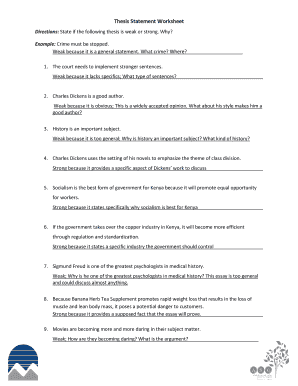
Practical instructions for preparing your ‘Thesis Statement Worksheet’ online
Are you fed up with the trouble of managing paperwork? Look no further than airSlate SignNow, the leading electronic signature solution for individuals and small to medium-sized businesses. Bid farewell to the labor-intensive process of printing and scanning documents. With airSlate SignNow, you can effortlessly complete and sign documents online. Utilize the powerful features embedded in this user-friendly and economical platform and transform your method of document management. Whether you need to authorize forms or collect electronic signatures, airSlate SignNow manages everything seamlessly, requiring just a few clicks.
Follow this comprehensive guide:
- Log into your account or register for a complimentary trial with our service.
- Click +Create to upload a file from your device, cloud storage, or our template library.
- Open your ‘Thesis Statement Worksheet’ in the editor.
- Click Me (Fill Out Now) to set up the document on your end.
- Add and assign fillable fields for others (if needed).
- Proceed with the Send Invite settings to solicit eSignatures from others.
- Download, print your version, or convert it into a reusable template.
No worries if you need to collaborate with your colleagues on your Thesis Statement Worksheet or send it for notarization—our solution has everything you require to complete such tasks. Create an account with airSlate SignNow today and elevate your document management to new levels!


