
Fill and Sign the Credit Check Authorization Letter Sample Form
Practical advice for finalizing your ‘Credit Check Authorization Letter Sample’ online
Are you fed up with the burden of handling paperwork? Search no further than airSlate SignNow, the leading eSignature platform for individuals and enterprises. Bid farewell to the tedious process of printing and scanning documents. With airSlate SignNow, you can easily finalize and sign documents online. Utilize the extensive features embedded in this user-friendly and affordable platform and transform your method of paperwork management. Whether you need to approve forms or gather electronic signatures, airSlate SignNow takes care of it all seamlessly, with just a few clicks.
Follow this detailed guide:
- Access your account or initiate a complimentary trial with our service.
- Click +Create to upload a file from your device, cloud storage, or our template collection.
- Open your ‘Credit Check Authorization Letter Sample’ in the editor.
- Click Me (Fill Out Now) to complete the form on your part.
- Add and designate fillable fields for other participants (if required).
- Proceed with the Send Invite settings to seek eSignatures from others.
- Download, print your copy, or convert it into a multi-usable template.
No need to worry if you need to work with your colleagues on your Credit Check Authorization Letter Sample or send it for notarization—our platform provides everything you need to accomplish such tasks. Register with airSlate SignNow today and elevate your document management to a new standard!
FAQs
-
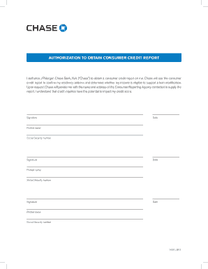
What is a Credit Check Authorization Letter Sample?
A Credit Check Authorization Letter Sample is a template used to grant permission to a lender or credit agency to access an individual's credit report. This letter ensures that the process is transparent and follows legal guidelines. Using a well-crafted sample can save time and help maintain professionalism in your communications.
-
How can airSlate SignNow help me with a Credit Check Authorization Letter Sample?
airSlate SignNow provides a user-friendly platform that allows you to easily create, edit, and eSign your Credit Check Authorization Letter Sample. With our intuitive interface, you can customize your letter to meet specific requirements, ensuring that it aligns with your business needs. Plus, you can send it directly to recipients for quick processing.
-
Is there a cost associated with using a Credit Check Authorization Letter Sample through airSlate SignNow?
Using a Credit Check Authorization Letter Sample on airSlate SignNow is cost-effective. We offer various pricing plans to fit different business needs, and our platform includes numerous templates, including credit check authorization letters, at no extra cost. This means you can save on both time and money while ensuring compliance.
-
What features does airSlate SignNow offer for creating a Credit Check Authorization Letter Sample?
airSlate SignNow offers several features for creating a Credit Check Authorization Letter Sample, including customizable templates, electronic signatures, and secure document storage. Our platform also supports team collaboration, allowing multiple users to work on the document simultaneously. This ensures that your letter is accurate and professionally presented before sending.
-
Can I integrate airSlate SignNow with other applications for managing Credit Check Authorization Letters?
Yes, airSlate SignNow offers seamless integrations with various applications, enabling you to manage your Credit Check Authorization Letters more efficiently. You can connect with tools like Google Drive, Salesforce, and more to streamline your workflow. This integration helps ensure that your documents are easily accessible and organized.
-
What are the benefits of using a Credit Check Authorization Letter Sample?
Utilizing a Credit Check Authorization Letter Sample can simplify the process of obtaining a credit report while ensuring compliance with legal requirements. It provides a clear and professional framework for communication between parties. Additionally, using airSlate SignNow enhances this process by allowing for quick eSigning and secure document handling.
-
How can I ensure my Credit Check Authorization Letter Sample is legally compliant?
To ensure your Credit Check Authorization Letter Sample is legally compliant, it's important to include specific information such as the recipient's details, the purpose of the request, and your consent for the credit check. airSlate SignNow provides guidance on creating legally sound documents, helping you adhere to necessary regulations and best practices.
Find out other credit check authorization letter sample form
- Close deals faster
- Improve productivity
- Delight customers
- Increase revenue
- Save time & money
- Reduce payment cycles
