Fill and Sign the Photosynthesis Graphic Organizer PDF Form
How it works
Open the document and fill out all its fields.
Apply your legally-binding eSignature.
Save and invite other recipients to sign it.
Rate template
4.4
Satisfied
47 votes
Next Generation Sequencing Application of DNA from FFPE Samples
Comprehensive Analysis of Your Stored Samples
1. Introduction
Formalin-Fixed Paraffin-Embedded (FFPE) samples are common
biological material for disease diagnoses and scientific research. This
technology allows samples to be stored for several years. It is however
challenging to get intact information from such samples, since severe
degradation, damage and molecular or biological modification could
appear during sample preparation. Although there are several kinds of
array-based methods for FFPE tissues, they are in general limited by
capacity and fixed targets. Next Generation Sequencing (NGS) for FFPE
samples will substantially facilitate our understanding of undefined
pathological mechanism and help to broaden our insights into biomedical
research.
As the premier scientific partner of scientists and researchers, BGI works
relentlessly in applying NGS to FFPE samples to unveil the information
concealed in FFPE samples.
2.2 Whole Genome Resequencing
Genomic DNA
Fragmentation
Randomly Fragment
Genomic DNA
Adapter Ligation
DNA Fragment
with Adapters
Clean Data Conversion Sequencing
(G)
Rate (%)
Fold (X)
Mapped
Rate (%)
Depth
(X)
Coverage
>=10X (%)
FFPE-1
212.28
194.21
91.49%
FFPE-2
130.71
120.18
91.94%
63.77
72.43%
25.17
88.47%
37.63
73.71%
24.84
FFPE-3
175.94
162.18
92.61%
92.18%
51.61
79.38%
37.35
Average
-
95.80%
-
91.87%
-
75.17%
29.12
Non-FFPEs*
-
92.29%
-
90%
-
85%
30
95%
Note:
[2] Clean data comes from raw data after filtering low quality reads and reads with adaptors.
DNA Cluster
[3] Conversion rate is the ratio between clean data and raw data.
[4] Sequencing fold is the ratio of clean data to the effective genome size.
[6] Depth is the ratio of clean data filtered out PCR duplication data to the effective genome
size.
2.1 Workflow of FFPE Sample Sequencing
Sequencing
Sequencing
Figure 2 WGRS Workflow
2.2.1 DNA Extraction and Library Construction
[7] Coverage ≥10× is the percent of effective genome size, which is covered by at least ten
reads.
2.3 Whole Exome Sequencing
Exon1
The QIAamp® DNA FFPE Tissue Kit (QIAGEN) with in-house modification was
used to extract total DNA from cervical tumor tissue FFPE samples. Although
DNA degradation existed, we successfully constructed WGRS libraries (data
not shown).
Bioinformatics
Analysis
Protease K Digestion
DNA Purification
and Isolation
Figure 1 Workflow of FFPE Sample Sequencing
WGRS libraries were assayed for sequencing using Illumina HiSeqTM 2000. The
effective average sequencing depth of each sample was 25~37×. As a measure
of how successful the sequencing of total DNA extracted from FFPE samples
was, we compared the conversion rate, mapped rate and coverage between the
non-FFPE and the FFPE samples as shown in Table 1. Results show that
FFPE-derived and non-FFPE-derived sequencing data have high conversion
rate (clean data/raw data), indicating the total DNA isolated with the extraction
kits tested was of sufficient quantity and quality for sequencing using Illumina
HiSeqTM 2000. In general, slightly more mapped rate and coverage were
detected in the non-FFPE samples than in FFPE samples, indicating that better
retention of the DNA during storage and total DNA extraction was achieved.
Exon2
Exon3
Genomic DNA
Wash
2.2.2 Sequencing Results
Paraffin Removal
Sample QC
Raw Data
(G)
[5] Mapped rate is the percent of clean data mapped to the effective genome size.
Among various NGS applications, Whole Genome Resequencing
(WGRS) and Whole Exome Sequencing (WES) are both commonly used
genomic research methods. Plenty of biomedical and drug R&D work have
already proved the substantial power of these NGS methods in locating
key disease related genes, profiling global mutations, biomarker
discovery, etc. There is a large source of useful data available on FFPE
samples. NGS is the premier choice for a comprehensive utilization of
these precious resources.
Library
Construction
Sample
[1] Non-FFPE* means normal sample without FFPE process, the data of non-FFPEs are
conservative empirical data based on BGI’s finished projects.
Amplification
2. Tests for FFPE Samples
FFPE Sample
Sections
Table 1 Comparison of WGRS Data Outcomes between FFPE and Non-FFPE* Samples
Exon4
Exon5
Exon6
Fragment
& Add Adapters
LM-PCR to Check Library Prep,
then Hybridize with Microarray
Background DNA
Target DNA
Elute
LM-PCR Amplification
Probes
qPCR Analysis:
QC of Capture
Success
Target
DNA
Sequencing
NimbleGen 2.1M Exome Array
Figure 3 WES Workflow
Next Generation Sequencing Application of DNA from FFPE Samples
Comprehensive Analysis of Your Stored Samples
2.3.1 DNA Extraction and Library Construction
The QIAamp® DNA FFPE Tissue Kit (QIAGEN) with in-house modification
was used to extract the total DNA from gastric tumor-adjacent tissue FFPE
samples, while the QIAamp® DNA mini Kit was used to extract total DNA
from matched Fresh Frozen (FF) samples. The degree of extracted DNA
degradation was evaluated by agarose gel electrophoresis. As expected,
analysis of DNA isolated from FF and FFPE tissues for the matched tumor
samples shows that genomic DNA from FF samples was of much higher
molecular weight and less degraded than that from FFPE tissues (Figure
4). We successfully constructed exome capture libraries from FFPE
sample using NimbleGen 2.1 M array, which was similar to FF sample
showed in Figure 5.
1 2 3 4
Table 2 Comparison of WES Data between FFPE and FF Samples
Samples
Raw
Data (M)
Mapped
Data (M)
Mapped
Rate (%)
FFPE
1419.41
1165.91
82.14
FF
1763.11
1490.43
84.53
Coverage
Mean Depth Coverage
of Target
of Target of Target
Region
Region (×) Region (%)
≥10× (%)
Capture
Specificity
(%)
18.61
93.31
65.52
65.22%
23.33
94.84
74.14
64.07%
Note:
[1] Mapped data represents data that was mapped to reference genome.
[2] Mapped rate is the percentage of raw data mapped with respect to the effective genome size.
[3] Mean depth of target region is the ratio of mapped data to the target region size.
[4] Coverage of target region is the percentage of target region covered by at least one read.
[5] Coverage of target region ≥10× is the percentage of target region covered by at least ten reads.
Figure 4 Total DNA Extracted from FF and FFPE Samples of
Gastric Paracancerous Tissue
[6] Capture specificity is the percentage of raw data mapped to the target region.
Lane 1: D2000 DNA marker;
Lane 2: Total DNA extraction from gastric paracancerous tissue FF
sample;
Lane 3: Total DNA extraction from gastric paracancerous tissue FFPE
sample;
Lane 4: λHind III DNA marker.
The figure shows that the DNA band from FFPE sample moved further
down compared to the band from FF sample, which indicates some
degradation.
1 2 3 4
Figure 5 Exome Capture Library from FF and FFPE Samples of
Gastric Paracancerous Tissue
Lane 1: D2000 DNA marker
Lane 2: Exome capture library constructed from FF sample
Lane 3: Exome capture library constructed from FFPE sample
Lane 4: 50 bp Ladder DNA marker
2.3.3 SNP Calling Results
Sequencing data was analyzed and evaluated for sequence coverage and
uniformity (Table 2), along with Single Nucleotide Polymorphism (SNP)
detection and concordance between the FF and FFPE samples (Figure 6 and
Figure 7). Concordance for normal SNPs (with both allele depth higher than 4×)
and high quality SNPs (with both allele depth higher than 20× and quality score
> 20) was approximately 98%, which indicated that we can get reliable genome
variation from FFPE samples that is comparable with FF samples.
Sample Requirement
Both the original FFPE samples and the DNA materials extracted are
acceptable. We recommend clients to extract DNA themselves.
FFPE Samples
Please make sure that each FFPE sample has no less than 20
sections, each of about 10-20 μm thick. About 100 μm sections are
required for DNA extraction each time. For example, if the FFPE
section has a thickness of 20 μm, 5 sections are needed for DNA
extraction. Please transfer tissue sections to clean centrifuge tubes by
clean nippers after section processing. To avoid cross contamination,
be sure to change different blades and nippers when transferring
different samples. BGI does not accept stained FFPE sections.
FFPE tissue sections provided by our collaborator are required to be
unstained; this can be sent under room temperature.
DNA Samples
• For Whole Genome Resequencing
Amount: 3×(N+1) μg; Concentration: ≥ 50 ng/μL; Purity: OD260/280=
1.8~2.0
N represents the number of library construction.
• For Whole Exome Sequencing
Amount: 6 μg; Concentration: ≥ 50 ng/μL; Purity: OD260/280 = 1.8~2.0
FFPE
FF
668
2.23%
28712
95.89%
FFPE
FF
562
158
7840
158
1.88%
1.94%
96.12%
1.94%
The figure shows the result of library construction with high-similarity
between FFPE sample and FF sample.
2.3.2 Sequencing Results
Figure 6 Concordance of ‘Normal’
SNPs between FF and FFPE Samples
WES libraries were assayed for sequencing using Illumina HiSeqTM 2000.
The mean depth of target region of each sample was 18.6× and 23.3×
respectively. To measure of how successful the sequencing of total DNA
extracted from FFPE samples was, we compared the mapped rate and
coverage between the FF and the FFPE samples as shown in Table 2.
Results show that sequencing data generated from FFPE and FF samples
had high mapping rate (mapped data / raw data), indicating that the total
DNA isolated with the extraction kits tested was of sufficient quantity and
quality for sequencing using Illumina HiSeqTM 2000. In general, slightly
more coverage was detected in the FF samples than in FFPE samples,
indicating that better retention of the DNA during storage and total DNA
extraction was achieved.
3. Conclusions
Figure 7 Concordance of ‘High Quality’
SNPs between FF and FFPE Samples
Most tumor tissue samples are preserved in the form of FFPE blocks, which in
general present several challenges, including variability of fixation methods,
diverse ages and store environment of samples, and several possible damages
that may occurs to the DNA during the FFPE process.
The results presented in this study demonstrate that DNA amplified from
degraded FFPE DNA can be successfully used for WGRS and WES studies
while maintaining acceptable levels of performance and allowing the
exploitation of FFPE samples by NGS technologies. These results enable the
use of a vast amount of FFPE samples available for biomedical researches and
applications.
Contact Us
China (Mainland)
BGI-Shenzhen, Beishan Industrial
Zone, Yantian District, Shenzhen,
518083, China
Tel: 400-706-6615
+86-755-25281960
Email: bgi-PGx@genomics.cn
www.genomics.cn
Asia Pacific(Hong Kong)
BGI Hong Kong Co. Limited, 16th Dai
Fu Street, Tai Po Industrial Estate, Tai
Po, Hong Kong
Tel: +852-3610-3510
Email: bgihk.enquiry@genomics.cn
www.bgisequence.com
Europe (Copenhagen)
Ole Maaløes Vej 3, DK-2200
Copenhagen N, Denmark
Tel: +45-7026 0806
Email: bgieurope@genomics.cn
www.bgisequence.com
North America (Boston)
BGI Americas Corporation, One
Broadway, 14th Floor, Cambridge, MA
02142, USA
Tel: +1-617-500-2741
Email: info@bgiamericas.com
www.bgiamericas.com
www.bgisequence.com
Valuable advice on preparing your ‘Photosynthesis Graphic Organizer Pdf’ digitally
Are you fed up with the inconvenience of handling paperwork? Look no further than airSlate SignNow, the leading electronic signature platform for individuals and small to medium-sized businesses. Bid farewell to the tedious process of printing and scanning documents. With airSlate SignNow, you can effortlessly finish and sign documents online. Take advantage of the robust features packed into this intuitive and cost-effective platform and transform your approach to document management. Whether you need to authorize forms or gather eSignatures, airSlate SignNow manages it all efficiently, requiring only a few clicks.
Follow this comprehensive guide:
Sign in to your account or register for a no-cost trial with our service.
Click +Create to upload a document from your device, cloud storage, or our template library.
Open your ‘Photosynthesis Graphic Organizer Pdf’ in the editor.
Click Me (Fill Out Now) to fill out the form on your end.
Add and designate fillable fields for others (if needed).
Continue with the Send Invite settings to solicit eSignatures from others.
Download, print your version, or convert it into a reusable template.
Don’t fret if you need to collaborate with your teammates on your Photosynthesis Graphic Organizer Pdf or send it for notarization—our solution has everything you need to accomplish these tasks. Create an account with airSlate SignNow today and elevate your document management to a higher level!
FAQs
Here is a list of the most common customer questions. If you can’t find an answer to your question, please don’t hesitate to reach out to us.
The Photosynthesis Graphic Organizer Pdf is a visual tool designed to help students and educators understand the process of photosynthesis. This PDF format organizer allows users to neatly outline the stages of photosynthesis, making it easier to study and retain information. By using this resource, learners can enhance their grasp of complex biological concepts.
You can easily access the Photosynthesis Graphic Organizer Pdf by visiting our website and downloading it directly. Simply navigate to the resources section, where you will find the PDF available for download. This ensures that you can start using the organizer immediately for your educational needs.
Yes, the Photosynthesis Graphic Organizer Pdf is available for free download. We believe in providing valuable educational resources without any cost, allowing students and educators to benefit from our tools without worrying about fees. Start enhancing your learning experience today!
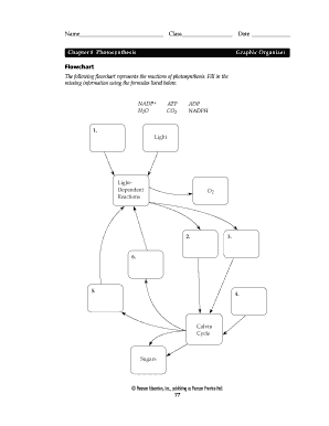
The Photosynthesis Graphic Organizer Pdf features a clear layout that highlights key components of the photosynthesis process, such as light-dependent reactions and the Calvin cycle. This user-friendly design makes it easy for students to fill in details and visualize the information. It's an excellent study aid for both classroom and home use.
The Photosynthesis Graphic Organizer Pdf benefits students by simplifying complex information into an easy-to-understand format. It encourages active learning as students can engage with the material by filling in the organizer. This enhances retention and comprehension of photosynthesis, which is crucial for their science education.
Absolutely! The Photosynthesis Graphic Organizer Pdf can seamlessly integrate with other teaching materials such as textbooks, presentations, and lab activities. Educators can use it alongside various resources to create a comprehensive lesson plan, enhancing the overall learning experience for students.
While the Photosynthesis Graphic Organizer Pdf is designed as a standard template, you can add your notes or specific instructions to tailor it to your classroom needs. This customization allows you to focus on particular aspects of photosynthesis that you want your students to emphasize, making it even more effective.
Related links photosynthesis graphic organizer pdf form
We use cookies to improve security, personalize the user experience, enhance our marketing activities (including cooperating with our 3rd party partners) and for other business use. Click here to read our Cookie Policy. By clicking “Accept“ you agree to the use of cookies.... Read moreRead less